Posts

Showing posts with the label Interventional Neuroradiology Specialist in North India

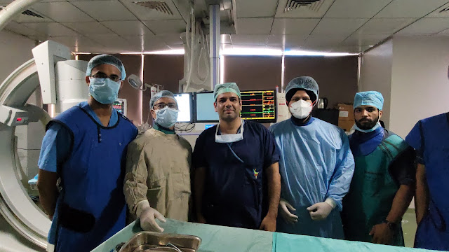
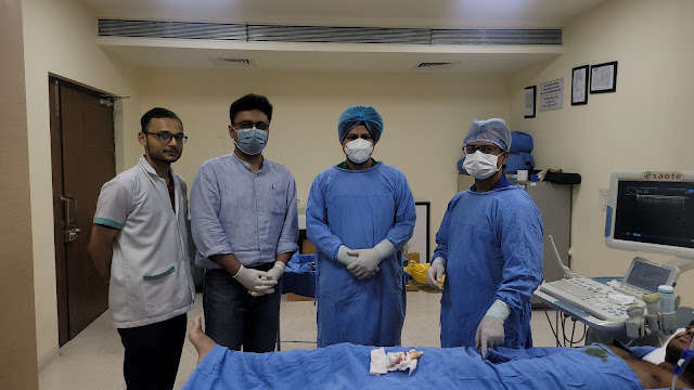
Multiple- Five Metastases Microwave Ablation

Image
Microwave ablation is a type of thermal ablation used to treat cancer in interventional radiology. To create tissue-heating effects, MWA uses electromagnetic waves in the microwave energy band (300 MHz to 300 GHz). Frictional heating is caused by the oscillation of polar molecules, which leads to tissue necrosis in solid tumours. It's typically utilised to treat and/or relieve the symptoms of solid tumours in patients who aren't surgical candidates. While some liver tumours can be surgically removed, the vast majority are inoperable and must be treated with other methods. Ablation (tissue destruction) is one such method. It is a surgical treatment that has traditionally been conducted using a variety of techniques, including Request for Applications (RFA) (Radiofrequency Ablation). People with liver metastases may benefit from ablation therapy, which can help lower the chance of cancer recurrence. Although ablation therapies do not eradicate tumours, they can result in complet...

Orbital Cancer Biopsy Carcinoma

Image
Adenoid cystic carcinoma is a rare epithelial-based malignant tumour that mainly arises from the salivary glands. A direct or perineural spread from a lacrimal gland or sinonasal source can cause orbital involvement. Primary orbital adenoid cystic carcinoma without lacrimal gland involvement is uncommon.  The earliest symptoms of AdCC are determined by the tumour's location. Early lesions of the salivary glands may present as painless, slow-growing lumps beneath the normal mouth lining or facial skin. Because there are numerous salivary glands beneath the mucosal lining of the mouth, throat, and sinuses, lumps in these areas could be the result of a tumour of this sort. Other signs and symptoms could include: ·  A mass on the roof of the mouth, under the tongue, or in the gums ·  A change in the mouth's lining. ·  Upper jaw, palate, face, and tongue numbness ·  Swallowing difficulties ·  Hoarseness ·  Pain is dull. ·  A lump or nodule in the earl...

Carotid Stenting With and Without Filter

Image
Carotid artery stenting is an endovascular technique that involves placing a stent into the carotid artery lumen to cure stenosis and reduce the risk of stroke. When carotid endarterectomy is deemed too dangerous, it is used to treat the narrowing of the carotid artery in high-risk patients. Traditional treatment for high-grade asymptomatic and symptomatic carotid artery stenosis has been carotid endarterectomy (CEA). Carotid endarterectomy is a procedure that involves exposing the carotid artery and removing plaque from the carotid bulb and proximal internal carotid artery through a neck incision. Minimally invasive procedures have evolved in vascular surgery, as they have in many other surgical disciplines, throughout time. This activity explains carotid artery stenting, including indications, contraindications, and possible problems.  Angioplasty and stenting of the carotid arteries may be effective stroke treatments or stroke prevention options. if:- ü  You have a 70% blo...

Cervical Lymph Node Multiple Biopsies In An Adolescent

Image
Cervical nodes that are swollen are rarely a symptom of malignancy.  However, non-painful enlargement of one or more lymph nodes, particularly those in the neck, is a crucial warning sign of lymphoma, including Hodgkin lymphoma (HL) and non-Hodgkin lymphoma (NHL) (NHL) Lymph nodes swell when viruses, dangerous bacteria, and damaged cells are trapped inside, and lymphocytes, the white blood cells that fight infection, strive to eliminate them. Swollen lymph nodes, on the other hand, can be an indication of malignancy, including lymphoma, a type of blood cancer. An incision is made over the swollen lymph node, and the node is dissected out of its surrounds with care to tie off or cauterise veins and lymphatic channels linked to it while under general anaesthesia. After that, the lymph node is transported to the lab for examination. Many infectious, autoimmune, metabolic, and malignant diseases affect the lymph nodes, which are an important part of the body's immune system. The ...

TACE For Large Arterio Portal Shunting

Image
Shunts between the portal venous and hepatic artery systems within the liver are known as intrahepatic arterioportal shunts. They may be the cause of reversible portal hypertension. The size and other underlying diseases will determine the clinical characteristics. Small shunts may go symptomless.  ·  The following are the several types of intrahepatic vascular shunts: ·  Transtumoural shunt occurs in hepatocellular carcinoma and, to a lesser extent, hepatic hemangioma due to improper communication between the tumour's feeding artery and draining vein, resulting in enhanced vascularity surrounding the tumour seen as temporary hepatic attenuation abnormalities (THAD) ·  The portal vein may exhibit an early increase on a dynamic arterial scan without the splenic and superior mesenteric veins, which are its main tributaries. Radiographic characteristics In general, the symptoms are comparable to those of cerebral infarction in any other location. As a result, these char...

Basilar Top Aneurysm

Image
Aneurysms of the basilar trunk artery are extremely rare, accounting for only 2.1 percent of all intracranial aneurysms. They are most commonly diagnosed in persons over 60, have a slight male predominance, and are linked to high morbidity and mortality. A type of posterior circulation cerebral aneurysm that accounts for just a small percentage of all aneurysms, with the majority occurring in the anterior circulation. Cerebral aneurysms are commonly found near bifurcations, such as this one, where the posterior cerebral artery is immediately posterior.   Ruptured basilar tip aneurysms can cause deadly subarachnoid hemorrhage (SAH), with a fatality rate of up to 23%. A big basilar artery aneurysm presenting as a stroke is extremely uncommon. The consequences and prognostic implications of this disorder necessitate examination and recognition.  Fusiform aneurysms are most common in severely atherosclerotic basilar arteries, and atherosclerotic disease is most often the cau...

LIVER ABSCESS DRAINAGE

Image
A pus-filled patch of fluid within the liver is known as a pyogenic hepatic abscess. Pyogenic denotes the production of pus. A liver abscess can arise from a number of various causes, including a blood infection, an abdominal infection, or an infected abdominal injury. An abscess in the liver is a serious infection. It has the potential to cause major issues and even death. It can cause tissue damage in the spot where it is discovered. Fever, discomfort, nausea, diarrhea, and loss of appetite are all possible symptoms. The whole first treatment for an amebic liver abscess is amebicidal medicines. The medication of choice is metronidazole. The size of the abscess is a key determinant in deciding how well medical therapy works. Other treatment options include percutaneous needle aspiration and/or catheter drainage.  The type of abscess that occurs is influenced by one's age. People in their 40s and 60s are more likely to have a liver abscess that is not caused by trauma.  ...

Vertebral Hemangioma Alcohol Ablation 

Image
Vertebral hemangiomas are benign vascular tumours of the spine that affect around ten percent of the population. They are frequently asymptomatic and are discovered by chance due to their distinctive features on MRI. If a hemangioma has unrestrained bleeding, it might be severe. For vertebral hemangiomas , alcohol ablation is an effectual treatment choice. Hemangioma discomfort can be effectively treated with radiation therapy. Surgery, radiotherapy into hemangiomatous vertebrae have all been used to treat vertebral hemangiomas. Vertebral hemangiomas appear as lucent patches separated by bony trabeculae on CT scans. V ertebral hemangiomas are congenital vascular abnormalities, not actual tumours. Using CT-guided alcohol ablation, about 85 percent of symptomatic vertebral hemangiomas can be improved significantly.    The non-surgical treatment for hypertrophic cardiomyopathy is alcohol septal ablation. The diagnosis of a vertebral hemangioma is critical and, in some situations,...

Acute Stroke Mechanical Thrombectomy

Image
Mechanical thrombectomy is indicated for patients who have an acute ischemic stroke caused by a large artery occlusion in the anterior circulation and can be treated within 24 hours of last known good health (ie, at neurologic baseline), regardless of whether they have received intravenous thrombolytic therapy for the same. Mechanical thrombectomy is a minimally invasive technique in which an interventional radiologist removes a clot from a patient's artery using specialised equipment. The doctor uses fluoroscopy, or continuous x-ray, to guide devices through the patient's arteries to the clot, which is then extracted in one piece. For patients with ischemic stroke (IS) who have a proximal major cerebral artery occlusion within 6 hours of onset of symptoms, mechanical thrombectomy (MT) is the standard-of-care treatment. In patients with anterior circulation ELVO and an NIHSS score of 6 [class I, level A], thrombectomy is recommended. When coupled with disabling symptoms [class...

Interventional Neuroradiology Specialist in North India

Image
  It started in 2012 with the effort of family members, friends and colleagues of Dr. Sandeep Sharma through visiting practice of Interventional Neuroradiology in north India . The interventional radiology and interventional neuroradiology are the latest and most advanced field in the medical science. However limited number of hospitals have this service and even fewer experts are available. Nevertheless, these services can have dramatic results and may cure many diseases in both elective and emergency situations. Also, the patient so treated has no stitches or scar and can be discharged almost immediately. These days care procedure can provided miraculous cures in health care.