Posts

Showing posts with the label Interventional Radiologist in Panchkula

How to Understand Varicose Veins Treatment in India

Image
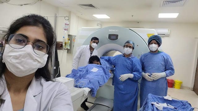
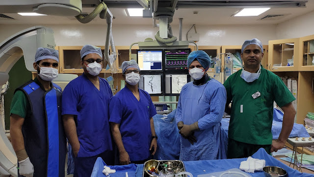
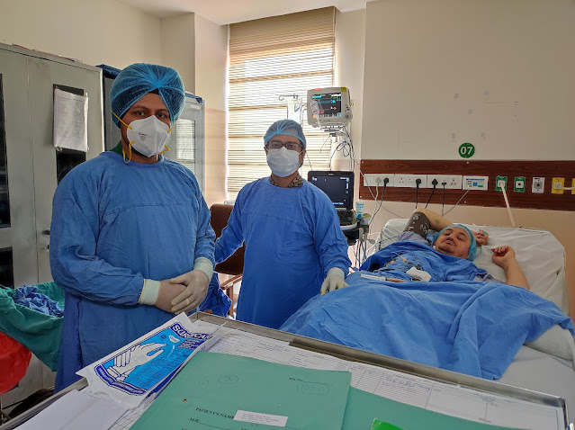
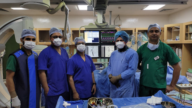
  Though India is a hub of varicose veins treatment, patients from these services can select from various advanced treatments. A few are: Laser Therapy (EVLT):  It is a minimally invasive procedure in which a beam of laser light is directed along the vein’s path it closing off the vein. Radiofrequency Ablation (RFA):  As the name suggests, the procedure uses radiofrequency energy to heat and close off very large veins. Sclerotherapy:  When it comes to varicose veins treatment in the lower limbs, the so-called foam sclerotherapy is the most common approach. The procedure involves injecting foam under ultrasound guidance per our standard operating procedure. Microphlebectomy:  An incision of about 1 to 3 mm is made through the skin where the smallest varicose veins are located, and they are pulled out from there. These treatments are performed by highly skilled vascular surgeons and interventional radiologists across the country. Top Doctors for Varicose Veins Tre...

Middle Meningeal Artery Embolization for Chronic SDH Post-Carotid Stenting | IRFACILITIES

Image
A patient with a history of chronic subdural hematoma (SDH) underwent carotid artery stenting, necessitating the initiation of dual antiplatelet therapy. Following this intervention, the patient developed an acute SDH, a known complication when antiplatelets are introduced in patients with pre-existing subdural collections. Given the risk of worsening hematoma due to antiplatelet therapy, a middle meningeal artery (MMA) embolization was performed. This minimally invasive procedure, which targets the vessels supplying the subdural space, was chosen to stabilize the chronic SDH and reduce the risk of further hemorrhage without discontinuing the necessary antiplatelet therapy. MMA embolization proved successful in managing the chronic SDH, mitigating the need for more invasive surgical interventions such as craniotomy, and allowing the patient to safely continue antiplatelet therapy for the recently placed carotid stent. The patient’s clinical course was favorable, with no further ep...

Vertebral Spine Biopsy

Image
A definitive tissue diagnosis of a vertebral lesion can be made with a minimally invasive, safe, and accurate method called CT-guided percutaneous vertebral biopsy . The most effective substitute for a surgical biopsy is frequently a CT-guided vertebral biopsy. The identification of spinal lesions is mostly dependent on magnetic resonance imaging (MRI). Even while recent advances in MRI now make it possible to recognize and suspect the nature of vertebral lesions and positron emission tomography-computed tomography (PET-CT) provides data on lesion metabolism, a biopsy is still required in the majority of instances. Both open surgery and a less invasive (percutaneous imaging-guided) procedure can be used to confirm the histopathology of a vertebral lesion. An open surgical biopsy of a vertebral lesion has a high risk of morbidity, the potential for contamination of nearby tissue planes, and potential postoperative problems. Under the direction of computed tomography, a percutaneous imag...

CT Guided Small Lung Nodule Biopsy

Image
CT-guided transthoracic needle biopsy is a simple and safe procedure that has a diagnostic accuracy of 64–97 percent for pulmonary lesions (5-8). As a result, one of the most common approaches for diagnosing the nature of pulmonary lesions is to use this process. For precise treatment of advanced non-small-cell lung cancer, molecular investigation of driver mutations is strongly suggested (NSCLC) CT-guided lung biopsy is a relatively safe and well-tolerated treatment. Some patients may endure substantial and long-term discomfort as a result of the treatment, and they should be informed about this beforehand. The doctor will put the needle through the skin, advance it to the site of the nodule, and remove tissue samples using imaging guidance. For a comprehensive analysis, several samples may be required. Following the collection of biopsies, a pathologist will rapidly review the samples to ensure that they are in good condition. When imaging findings indicate stability or when consist...

CA Larynx With Lingual Artery Aneurysm Glue Embolisation

Image
A rare condition called postoperative lingual artery pseudoaneurysm coupled with bleeding is typically observed following laryngectomy or tonsillectomy. This study describes the successful use of catheter-directed glue-embolization to treat a symptomatic lingual artery pseudoaneurysm that had developed following the recurrent base of tongue surgeries. Damage to the mouth's floor, particularly iatrogenic trauma from surgical operations, might result in a false aneurysm. This article will detail a case of a lingual artery pseudoaneurysm that developed after tonsillectomy. After a tonsillectomy, lingual artery pseudoaneurysms can develop within a few hours. Endovascular intervention is an effective and low-morbidity alternative to surgery for the treatment of such an aneurysm, and angiography offers the diagnosis. Platinum coil endovascular embolization is a successful technique for managing bleeding and avoiding surgical intervention.  Intervention in the ECA should be a skill set fo...

Five Metastases Microwave Ablation

Image
Microwave ablation i s a technique for heating and killing cancer cells using high-frequency microwave energy. It's a treatment for certain types of big lung cancer. It has the ability to eliminate a tumour that is obstructing the airway. This makes it easier for you to breathe. Ablation is a treatment for liver tumours that kills them without removing them. When surgery is not an option, these procedures can be employed in patients with a few tiny tumours (often because of poor health or reduced liver function). In most cases, hepatic percutaneous microwave ablation (MWA) is conducted under conscious sedation. Despite this, numerous patients complained of discomfort throughout the operation. How long does it take to ablate a tumour? The needle warms the tumour, causing it to die. This can take a long time. It could take anywhere from 30 minutes to several hours to complete. Radiofrequency ablation (RFA) and microwave ablation (MWA) both employ radio waves to burn and eliminate neu...

Chemical Lumbar Sympathectomy

Image
The sympathetic nerves that run along the front of the backbone, near to but outside the spinal column, are injected with a chemical sympathectomy. Lumbar sympathectomy is used to treat leg and foot pain caused by a lack of blood supply, as well as to aid in the healing of leg and foot ulcers. To cure excessive perspiration in the foot and to reduce leg and back pain caused by spinal canal stenosis.  Lumbar Sympathectomy.  ü  A collection of nerves in your lower back is known as the lumbar sympathetic chain. The blood supply to the legs is controlled by these nerves. ü  A lumbar sympathectomy is a technique in which the sympathetic nerves are blocked and the blood supply to the skin in the leg is increased. This can help you feel better and heal faster. ü  Phenol is a drug that is commonly injected and can provide pain relief for up to 9 months. ü  Lumbar sympathectomy is used to treat leg and foot pain caused by a lack of blood flow; to aid in the healing...

Coiling Or Clipping In Brain Aneurysms

Image
The coil causes aneurysm thrombosis and is left in the aneurysm indefinitely. Surgical clipping needs open surgery and is performed under general anesthesia. To visualize the aneurysm, the brain is slowly retracted. To stop blood flow into the aneurysm, a tiny clip is inserted across its neck. A small metal clip is used to halt blood flow into the aneurysm during microsurgical clipping. To reach the aneurysm in the brain, a craniotomy is performed to make an opening in the skull. The clip is placed on the aneurysm's neck (opening) to impede blood flow, and it stays inside the brain. The clip functions similarly to a miniature coil-spring clothespin, with the blades remaining tightly closed until pressure is applied to open them. The titanium clips are permanently attached to the artery. In terms of mortality, re-bleeding, and re-treatments, neurosurgical cutting produces better results. In terms of post-operative problems, good outcomes, and rehabilitation, endovascular coiling is...

Trans Splenic Pancreatic Mass FNAC

Image
Endoscopic ultrasonography-guided fine-needle aspiration cytology (EUS-FNAC) is a minimally invasive procedure that is commonly used to assess deep-seated benign and malignant tumours. The utility of cytological samples in lymphoma diagnosis, on the other hand, is still debatable. Real-time puncture, reduced risk of complications due to the needle's proximity to the lesion, and the ability to sample small lesions that would be difficult to sample using other methods are all advantages of EUS over other imaging modalities. Finally, EUS provides access to deep-seated lesions, which is difficult to achieve with conventional methods. With an overall accuracy of 65 per cent to 100 per cent, EUS allows for sampling of mediastinal, retroperitoneal, and per gastrointestinal lymph nodes. Fine-needle aspiration biopsy guided by endoscopic ultrasonography. Through the mouth and into the oesophagus, an endoscope with an ultrasonography probe and a biopsy needle is introduced. The probe sends s...

Orbital Cancer Biopsy Carcinoma

Image
Adenoid cystic carcinoma is a rare epithelial-based malignant tumour that mainly arises from the salivary glands. A direct or perineural spread from a lacrimal gland or sinonasal source can cause orbital involvement. Primary orbital adenoid cystic carcinoma without lacrimal gland involvement is uncommon.  The earliest symptoms of AdCC are determined by the tumour's location. Early lesions of the salivary glands may present as painless, slow-growing lumps beneath the normal mouth lining or facial skin. Because there are numerous salivary glands beneath the mucosal lining of the mouth, throat, and sinuses, lumps in these areas could be the result of a tumour of this sort. Other signs and symptoms could include: ·  A mass on the roof of the mouth, under the tongue, or in the gums ·  A change in the mouth's lining. ·  Upper jaw, palate, face, and tongue numbness ·  Swallowing difficulties ·  Hoarseness ·  Pain is dull. ·  A lump or nodule in the earl...