Posts

Showing posts from June, 2022

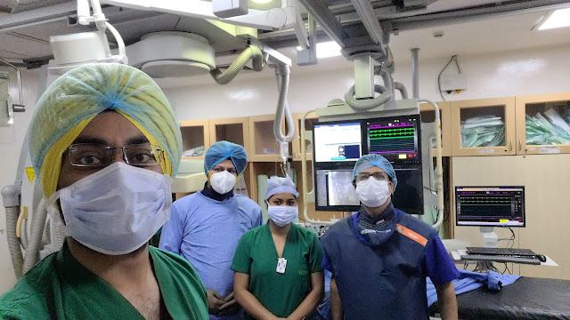
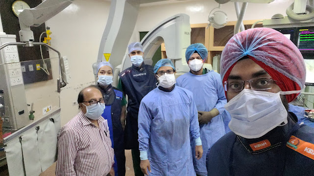
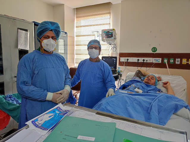
Subclavian Artery Stenting

Image
Due to the short- and long-term consequences, as well as patency difficulties, treatment for subclavian artery blockage is still up for debate. We present a rare instance of bilateral internal carotid artery occlusion and coupled proximal segmental occlusion of the left subclavian artery and subclavian steal phenomenon. Depending on the severity of the stenosis, the patient may exhibit symptoms or be asymptomatic. Upper extremity claudication, vertigo, diplopia, or syncope are some of the symptoms. A complete investigation using ultrasound Doppler, magnetic resonance angiogram (MRA), and computed tomography angiography (CTA) is undertaken to delineate the diagnosis when a blood pressure differential in the upper arms is greater than 15 mmHg. The management strategy involves medical care, endovascular therapy, and lifestyle changes, depending on the extent of the clinical manifestation. Endovascular stenting and angioplasty should be used as initial treatments for symptomatic subclavia

CA Larynx With Lingual Artery Aneurysm Glue Embolisation

Image
A rare condition called postoperative lingual artery pseudoaneurysm coupled with bleeding is typically observed following laryngectomy or tonsillectomy. This study describes the successful use of catheter-directed glue-embolization to treat a symptomatic lingual artery pseudoaneurysm that had developed following the recurrent base of tongue surgeries. Damage to the mouth's floor, particularly iatrogenic trauma from surgical operations, might result in a false aneurysm. This article will detail a case of a lingual artery pseudoaneurysm that developed after tonsillectomy. After a tonsillectomy, lingual artery pseudoaneurysms can develop within a few hours. Endovascular intervention is an effective and low-morbidity alternative to surgery for the treatment of such an aneurysm, and angiography offers the diagnosis. Platinum coil endovascular embolization is a successful technique for managing bleeding and avoiding surgical intervention.  Intervention in the ECA should be a skill set fo

Five Metastases Microwave Ablation

Image
Microwave ablation i s a technique for heating and killing cancer cells using high-frequency microwave energy. It's a treatment for certain types of big lung cancer. It has the ability to eliminate a tumour that is obstructing the airway. This makes it easier for you to breathe. Ablation is a treatment for liver tumours that kills them without removing them. When surgery is not an option, these procedures can be employed in patients with a few tiny tumours (often because of poor health or reduced liver function). In most cases, hepatic percutaneous microwave ablation (MWA) is conducted under conscious sedation. Despite this, numerous patients complained of discomfort throughout the operation. How long does it take to ablate a tumour? The needle warms the tumour, causing it to die. This can take a long time. It could take anywhere from 30 minutes to several hours to complete. Radiofrequency ablation (RFA) and microwave ablation (MWA) both employ radio waves to burn and eliminate neu

Chemical Lumbar Sympathectomy

Image
The sympathetic nerves that run along the front of the backbone, near to but outside the spinal column, are injected with a chemical sympathectomy. Lumbar sympathectomy is used to treat leg and foot pain caused by a lack of blood supply, as well as to aid in the healing of leg and foot ulcers. To cure excessive perspiration in the foot and to reduce leg and back pain caused by spinal canal stenosis.  Lumbar Sympathectomy.  ü  A collection of nerves in your lower back is known as the lumbar sympathetic chain. The blood supply to the legs is controlled by these nerves. ü  A lumbar sympathectomy is a technique in which the sympathetic nerves are blocked and the blood supply to the skin in the leg is increased. This can help you feel better and heal faster. ü  Phenol is a drug that is commonly injected and can provide pain relief for up to 9 months. ü  Lumbar sympathectomy is used to treat leg and foot pain caused by a lack of blood flow; to aid in the healing of leg and foot ulcers; to r

Carotid Artery Stenting

Image
For the treatment of carotid stenosis, carotid artery stenting (CAS) is a viable alternative to carotid endarterectomy (CEA). Carotid artery stenosis is a disabling condition that affects people of all ages. Because of their advanced age and many co-morbidities, the elderly are more susceptible to repeated strokes. The treatment choices for symptomatic carotid stenosis in the extremely elderly are the same as in younger patients, however, operation risk is increased. Practical Implications- Stenting of the carotid arteries is a common treatment option for atherosclerotic carotid artery stenosis. Carotid artery stenosis is a common cause of stroke in the elderly, and stroke outcomes are heavily impacted by age. After a thorough examination of the patient and the lesion, very elderly symptomatic patients should be studied and intervention considered. Background Carotid artery stenosis is a common cause of stroke in the elderly, and age has a significant impact on stroke outcomes. As the

Coiling Or Clipping In Brain Aneurysms

Image
The coil causes aneurysm thrombosis and is left in the aneurysm indefinitely. Surgical clipping needs open surgery and is performed under general anesthesia. To visualize the aneurysm, the brain is slowly retracted. To stop blood flow into the aneurysm, a tiny clip is inserted across its neck. A small metal clip is used to halt blood flow into the aneurysm during microsurgical clipping. To reach the aneurysm in the brain, a craniotomy is performed to make an opening in the skull. The clip is placed on the aneurysm's neck (opening) to impede blood flow, and it stays inside the brain. The clip functions similarly to a miniature coil-spring clothespin, with the blades remaining tightly closed until pressure is applied to open them. The titanium clips are permanently attached to the artery. In terms of mortality, re-bleeding, and re-treatments, neurosurgical cutting produces better results. In terms of post-operative problems, good outcomes, and rehabilitation, endovascular coiling is

Varicocele Embolization Using Sclerosant, Coils And Glue

Image
Varicocele is a condition in which the veins in the pampiniform plexus dilate abnormally due to blood reflux from the internal spermatic vein. Varicocele affects about 15-20 percent of adolescents and young guys, and up to 40-45 percent of infertile males have it. The cause of varicocele is unknown, but it is likely complex. The most prevalent cause of reflux in the internal spermatic vein is valve malfunction, which can be inherited or acquired. To divert blood flow away from a varicocele, a catheter (long, thin, hollow plastic tube) and imaging guidance are used to put small coils and/or liquid material in a blood vessel. It's less invasive than traditional surgery, and it can safely ease pain and swelling while also potentially improving sperm quality. The aberrant testicular vein is subsequently blocked with little metal coils. The radiologist will remove the catheter when the treatment is completed and gently press on the entry site for a few minutes to prevent bleeding. It wi

Celiac Axis Block For Cancer Related Pain

Image
Pancreatic cancer causes severe abdominal pain, which can be relieved by a celiac plexus block. The celiac plexus nerves in the abdomen stop providing pain signals to the brain due to this sort of nerve damage. Some types of celiac plexus block relieve pain temporarily, while others provide long-term relief. Pancreatic cancer is a type of cancer that causes considerable pain in 50 to 70% of patients and is notoriously difficult to cure. Celiac plexus block (CPB) is a pain-relieving procedure that is thought to be both safe and effective. In individuals with inoperable pancreatic cancer, abdominal pain is a common symptom that is difficult to treat. Celiac plexus block (CPB) is a pain-relieving procedure that is both safe and effective. The nerve fibres that transmit pain from the abdomen to the brain are chemically destroyed. In patients with inoperable pancreatic cancer, we looked for studies that compared CPB to routine analgesic therapy. The primary consequence of pain, as measured

Bilateral Varicocele Embolization

Image
Varicoceles are not life-threatening, however, they can be linked to serious illnesses in rare cases. If a varicocele arises on the right side of the abdomen rather than the left, for example, make sure there is no mass or other abnormalities in the abdomen that could be causing it. Blood is transported back to the heart via veins throughout the body. They usually feature valves to keep the blood flowing in the right way. When the valves in the testicular vein don't work properly, blood can pool in the scrotum, resulting in a varicocele. They commonly happen on the left side, and they're caused by the testicular vein's path through the abdomen. Varicocele is a frequent condition that is not hazardous. In fact, a varicocele affects 15% of all adult men. Many men's varicocele will go undiscovered for the rest of their lives or may create no complications at all. Varicoceles affect about 20% of teens, therefore a portion of them will most likely resolve on their own. The

Varicose veins

Image
Varicose veins are veins that are twisted and swollen. Varicose veins can develop near the skin's surface (superficial veins). Varicose veins are most typically found in the legs. Because standing and walking raise the pressure in the lower body's veins, this is the case. Varicose veins are gentle.   The cause of this illness is unknown. Varicose veins are a cosmetic concern for many people who have no symptoms. They might produce agonizing pain and discomfort, or they can indicate an underlying circulation condition. Compression stockings, exercise, and operations to seal or eliminate the veins are all options for treatment. Varicose veins major cause - Increased blood pressure in the veins causes varicose veins. Varicose veins develop in the veins near the skin's surface (superficial). One-way valves in the veins direct blood flow towards the heart. Blood can gather in the veins if the valves become weaker or broken. Varicose and spider veins do not disappear on their ow

Multiple- Five Metastases Microwave Ablation

Image
Microwave ablation is a type of thermal ablation used to treat cancer in interventional radiology. To create tissue-heating effects, MWA uses electromagnetic waves in the microwave energy band (300 MHz to 300 GHz). Frictional heating is caused by the oscillation of polar molecules, which leads to tissue necrosis in solid tumours. It's typically utilised to treat and/or relieve the symptoms of solid tumours in patients who aren't surgical candidates. While some liver tumours can be surgically removed, the vast majority are inoperable and must be treated with other methods. Ablation (tissue destruction) is one such method. It is a surgical treatment that has traditionally been conducted using a variety of techniques, including Request for Applications (RFA) (Radiofrequency Ablation). People with liver metastases may benefit from ablation therapy, which can help lower the chance of cancer recurrence. Although ablation therapies do not eradicate tumours, they can result in complet

R MCA Stroke Thrombectomy

Image
Patients with ischemic stroke (IS) who have a proximal major cerebral artery occlusion within 6 hours of the onset of symptoms are treated with mechanical thrombectomy (MT). If the patient qualifies, IV tissue plasminogen activator (TPA) and thrombectomy are the treatments for an MCA stroke. In patients with anterior circulation ELVO and an NIHSS score of 3, thrombectomy is recommended. In addition to changes owing to direct tissue damage to the insula and basal ganglia, a right MCA stroke may impair the feeling of pleasurable emotions via affecting brain activity in limbic and paralimbic regions far from the location of direct damage. The fast onset of a focused neurologic deficit caused by a brain infarction or ischemia in the territory supplied by the MCA is known as an MCA stroke. The MCA is the largest cerebral artery by far, and it is the artery most usually injured by a stroke.  The MCA's principal job is to provide oxygenated blood to specific areas of the brain parenchym

LIVER ABSCESS DRAINAGE

Image
A pus-filled patch of fluid within the liver is known as a pyrogenic hepatic abscess. Pyogenic denotes the production of pus. A liver abscess can arise from a number of various causes, including a blood infection, an abdominal infection, or an infected abdominal injury. An abscess in the liver is a serious infection. It has the potential to cause major issues and even death. It can cause tissue damage in the spot where it is discovered. Fever, discomfort, nausea, diarrhoea, and loss of appetite are all possible symptoms. The whole first treatment for an amebic liver abscess is amebicidal medicines. The medication of choice is metronidazeole. The size of the abscess is a key determinant in deciding how well medical therapy works. Other treatment options include percutaneous needle aspiration and/or catheter drainage.  The type of abscess that occurs is influenced by one's age. People in their 40s and 60s are more likely to have a liver abscess that is not caused by trauma.  The foll

Trans Splenic Pancreatic Mass FNAC

Image
Endoscopic ultrasonography-guided fine-needle aspiration cytology (EUS-FNAC) is a minimally invasive procedure that is commonly used to assess deep-seated benign and malignant tumours. The utility of cytological samples in lymphoma diagnosis, on the other hand, is still debatable. Real-time puncture, reduced risk of complications due to the needle's proximity to the lesion, and the ability to sample small lesions that would be difficult to sample using other methods are all advantages of EUS over other imaging modalities. Finally, EUS provides access to deep-seated lesions, which is difficult to achieve with conventional methods. With an overall accuracy of 65 per cent to 100 per cent, EUS allows for sampling of mediastinal, retroperitoneal, and per gastrointestinal lymph nodes. Fine-needle aspiration biopsy guided by endoscopic ultrasonography. Through the mouth and into the oesophagus, an endoscope with an ultrasonography probe and a biopsy needle is introduced. The probe sends s