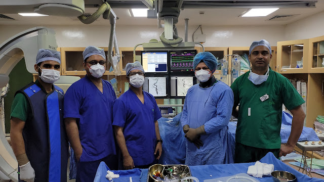
Trans Splenic Pancreatic Mass FNAC
Endoscopic ultrasonography-guided fine-needle aspiration cytology (EUS-FNAC) is a minimally invasive procedure that is commonly used to assess deep-seated benign and malignant tumours. The utility of cytological samples in lymphoma diagnosis, on the other hand, is still debatable. Real-time puncture, reduced risk of complications due to the needle's proximity to the lesion, and the ability to sample small lesions that would be difficult to sample using other methods are all advantages of EUS over other imaging modalities. Finally, EUS provides access to deep-seated lesions, which is difficult to achieve with conventional methods. With an overall accuracy of 65 per cent to 100 per cent, EUS allows for sampling of mediastinal, retroperitoneal, and per gastrointestinal lymph nodes.
Fine-needle aspiration biopsy guided by endoscopic ultrasonography. Through the mouth and into the oesophagus, an endoscope with an ultrasonography probe and a biopsy needle is introduced. The probe sends sound waves across the body, creating echoes that generate a sonogram of the lymph nodes around the oesophagus. The initial step in diagnosing a suspected pancreatic mass lesion is to use non-invasive imaging techniques such as computed tomography, ultrasonography, and magnetic resonance imaging. Tissue acquisition for characterization of a suspicious lesion is typically crucial in designing a personalised therapy approach once a suspect lesion has been identified. In addition to radiologic imaging, serum tumour markers, endoscopic retrograde cholangiopancreatography, Ultrasound-guided (USG) fine-needle aspiration cytology (FNAC), and endoscopic ultrasound-guided fine-needle aspiration (EUS-FNA) are currently used in the initial evaluation of a patient with a pancreatic mass lesion.
The accuracy, sensitivity, specificity, and positive predictive value of EUS-FNAC are all high. By overcoming the limitations of EUS-FNAC, it will become a valuable and trustworthy diagnostic tool for assessing pancreatic lesions accurately.
Signs and symptoms of pancreatic cancer in its early stages
Symptoms
· Back pain that originates in the abdomen.
· Appetite loss or unintentional weight loss
· Your skin and the whites of your eyes are turning yellow (jaundice)
· Stools in a light colour scheme.
· Urine with a dark colour.
· The skin is itchy.
· Newly diagnosed diabetes or diabetes that is becoming increasingly difficult to manage.
· Clots in the blood.
The procedure is likely to be utilised even more frequently in the future due to the documented high level of accuracy and broad margin of safety of radiologically guided needle biopsy.
More widespread use should result in better patient care by lowering the mortality, morbidity, and expense of unneeded surgery, as well as lowering the need for other diagnostic procedures. In the future, a guided needle biopsy is likely to have a broader range of uses. Follow-up biopsies of tumours for unique pathologic studies (for example, assays for special tissue receptors and chemotherapy drug levels from within the tumour can be obtained) and percutaneous needle treatment of certain diseases (for example, the direct percutaneous needle injection of alcohol into a tumour for ablation of that mas) are likely to be two of these future applications. Pancreatic growths, both benign and precancerous. Some pancreatic growths are benign (noncancerous), while others might progress to malignancy if left untreated (known as precancers). The pancreas has a tumour that is greater than 2 cm. It hasn't spread to lymph nodes or any other organs. Stage IIA: The tumour has grown to be more than 4 cm in diameter and has spread outside the pancreas.
Dr Sandeep Sharma is the Best doctor for Acute Stroke Mechanical Thrombectomy and the best radiologist. He is an expert in both suction retrieval and stent retrieval and uses both techniques in perfect combination


Comments
Post a Comment