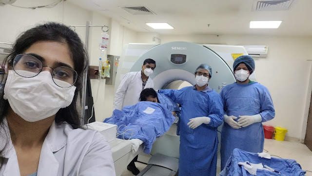
Vertebral Spine Biopsy
In comparison to standard blood tests, a slightly larger needle is utilized during the biopsy. The back of the hip's bone must be punctured with this larger needle. Following the biopsy, the bone hole will start to heal right away, and it is anticipated that full healing will have taken place in 1 to 2 weeks.
Technique
The CT-guided biopsy of vertebral lesions can be performed using a variety of approach-based approaches. These methods rely on a variety of variables, including the following: Location within the vertebrae, localized or diffuse lesion, location within the spinal column, such as the cervical, thoracic, or lumbar spine, vertebral plana, and accompanying soft tissue mass. Conscious sedation or local anesthetic is typically used during CT-guided vertebral biopsy procedures. Patients with comorbidities, those unable to lie prone, and young patients all necessitate general anesthetic.
The three Ps—"planning" for a successful biopsy, "positioning" the patient to achieve the ideal biopsy plane, and "protection" of vital vascular and neural structures in the trajectory of a biopsy needle—are the best way to express the key principles for a successful and safe CT-guided vertebral biopsy. Most CT-guided percutaneous vertebral biopsy procedures for the thoracic and lumbar vertebrae are carried out with the patient on their back. Patient posture for cervical vertebrae is influenced by the spot where the lesion is, how the biopsy is done, and the operator's competence level. The patient is positioned prone on the table for direct posterior and posterolateral approaches. The patient is positioned in the decubitus posture with the side of the lesion facing upwards for the direct lateral approach. In order to do the anterolateral approach, the patient is lying face down.
The numerous vertebral biopsy techniques include:-
Ø Transpedicular technique
Ø Posterolateral extravehicular technique
Ø Superior costotransverse technique
Ø Inferior costotransverse technique
Ø Pedicular biopsy
Ø Anterolateral or lateral technique
Ø Dual biopsy
Conclusion
The most effective alternative to open surgical vertebral biopsy for the accurate and safe outpatient diagnosis of the vertebral lesion is a CT-guided percutaneous vertebral biopsy. The rule of the three Ps, rigorous step-by-step planning, and customization of the approach based on the vertebral level involved are crucial to the procedure's effectiveness with the highest level of safety. The requirement for open surgical biopsy can be avoided by utilizing the discussed least invasive approach-based procedures.


Comments
Post a Comment